

Micro- & Nanoplastics
Light from the Crystal's Darkness: When Diamonds Trace Invisible Plastics
Corresponding author: Tatiana Kárová, [email protected]
Micro- and nanoplastic pollution is a major environmental concern and a growing subject of health research. Yet particles below 100 nanometers are difficult to detect and chemically characterize, especially in complex biological and environmental samples. This article examines whether nitrogen-vacancy (NV) centers in synthetic diamond could contribute to future nanoplastic sensing. We explain the principles of optically detected magnetic resonance and nanoscale nuclear magnetic resonance (nano-NMR), the production of near-surface NV centers, and the limitations imposed by surface noise. Existing studies have separately demonstrated nanoscale NMR, long-term tracking of model polymer particles with fluorescent nanodiamonds, and quantum sensing of free radicals in cells. Their combination into a direct, chemically specific nanoplastic detector, however, remains a future research goal. We conclude by discussing the engineering steps required for integrated and potentially field-deployable quantum sensing systems.
Article
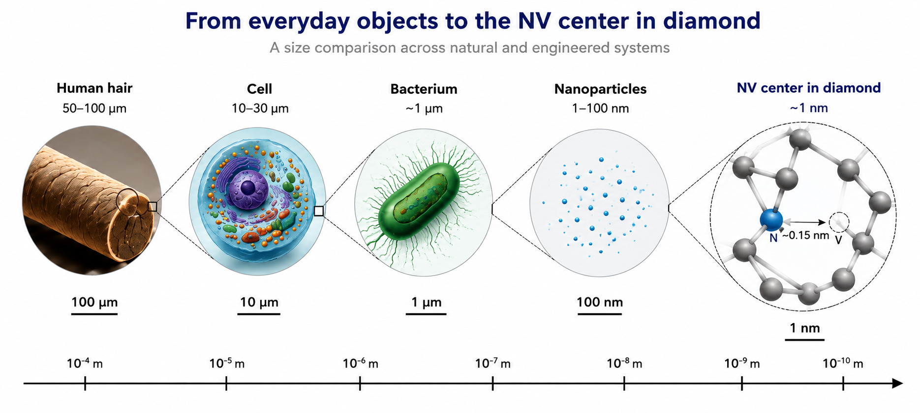
Finding and accurately identifying a plastic particle that is a thousand times thinner than a human hair, and located deep within living tissue or a natural water source, is a monumental challenge for contemporary science. Conventional optical methods commonly used in laboratories have rigid physical boundaries (Fig. 1). Quite simply, light cannot scatter properly on objects of such minuscule dimensions. Other analytical methods can identify the chemical composition of plastics, but they irreversibly destroy the sample in the process. Consequently, tracking the transport of nanoplastics in real time inside a living organism has long been exceptionally difficult. Surprisingly, a promising—though still largely experimental—solution is now emerging from the realm of quantum physics, utilizing technology hidden within laboratory-grown diamonds.
The Current State of Nanoplastic Detection and the Historical Context of NV Centers
The demand for reliable nanoplastic analysis is increasing. Clean laboratory conditions are no longer sufficient; scientists need to monitor pollution directly in ecosystems, and physicians are interested in observing how these tiny particles behave inside cells and organs [1]. However, once a plastic particle drops below the 100-nanometer threshold, conventional laboratory instruments essentially go blind.
Conventional Detection Technologies: Advantages and Limitations
Nanoplastic research uses a broad range of analytical approaches. Two methods that demonstrate current limitations and trade-offs are Raman spectroscopy and pyrolysis gas chromatography-mass spectrometry (Py-GC-MS) (Fig. 2).
- Raman spectroscopy works by illuminating a sample with a laser, causing the polymer molecules to vibrate. The scattered light carries their unique chemical fingerprint. The main advantage is that this method is non-destructive. However, conventional microscopes hit a fundamental limit called the diffraction limit: because of its wavelength, ordinary light cannot clearly resolve objects smaller than a few hundred nanometers. For nanoplastics, the signal drops off rapidly as the particles get smaller, meaning it practically vanishes for the tiniest fragments. Moreover, in biological samples, this weak signal is entirely drowned out by the natural autofluorescence of the surrounding tissue [1].
- Pyrolysis-GC-MS takes a different approach. The sample is literally vaporized at extreme temperatures in an oxygen-free environment, and the instrument analyzes the mass of the resulting molecular fragments. The advantage lies in its extreme sensitivity, which can detect trace amounts of a specific polymer. The overarching disadvantage, however, is its destructive nature. Because the material is permanently destroyed, its original size, shape, and biological context are completely lost, and the same particle cannot be followed over time [1].
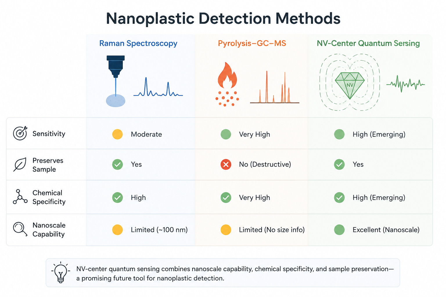
Scientists are therefore exploring complementary approaches, and one promising possibility involves nitrogen-vacancy centers in diamond.
A Brief History of NV Centers
The history of nitrogen-vacancy centers dates back to the 1960s, when researchers began investigating the unusual optical properties of radiation-damaged diamonds. A major breakthrough occurred in 1997, when a team at Chemnitz University of Technology demonstrated that the spin state of a single isolated defect could be detected at room temperature using confocal optical microscopy [2]. This milestone helped establish NV centers as a platform for quantum sensing and metrology. Around 2008, researchers demonstrated that individual NV centers could function as nanoscale magnetic-field sensors (Fig. 2). Several years later, NV centers were also developed into nanoscale thermometers [3]. More broadly, the possible use of quantum sensing to detect microplastics has recently begun to attract attention, although it remains an exploratory research direction [4].
The Physical Principles of NV Centers and Quantum Nanoplastic Sensing
The Quantum Structure of an "Artificial Atom"
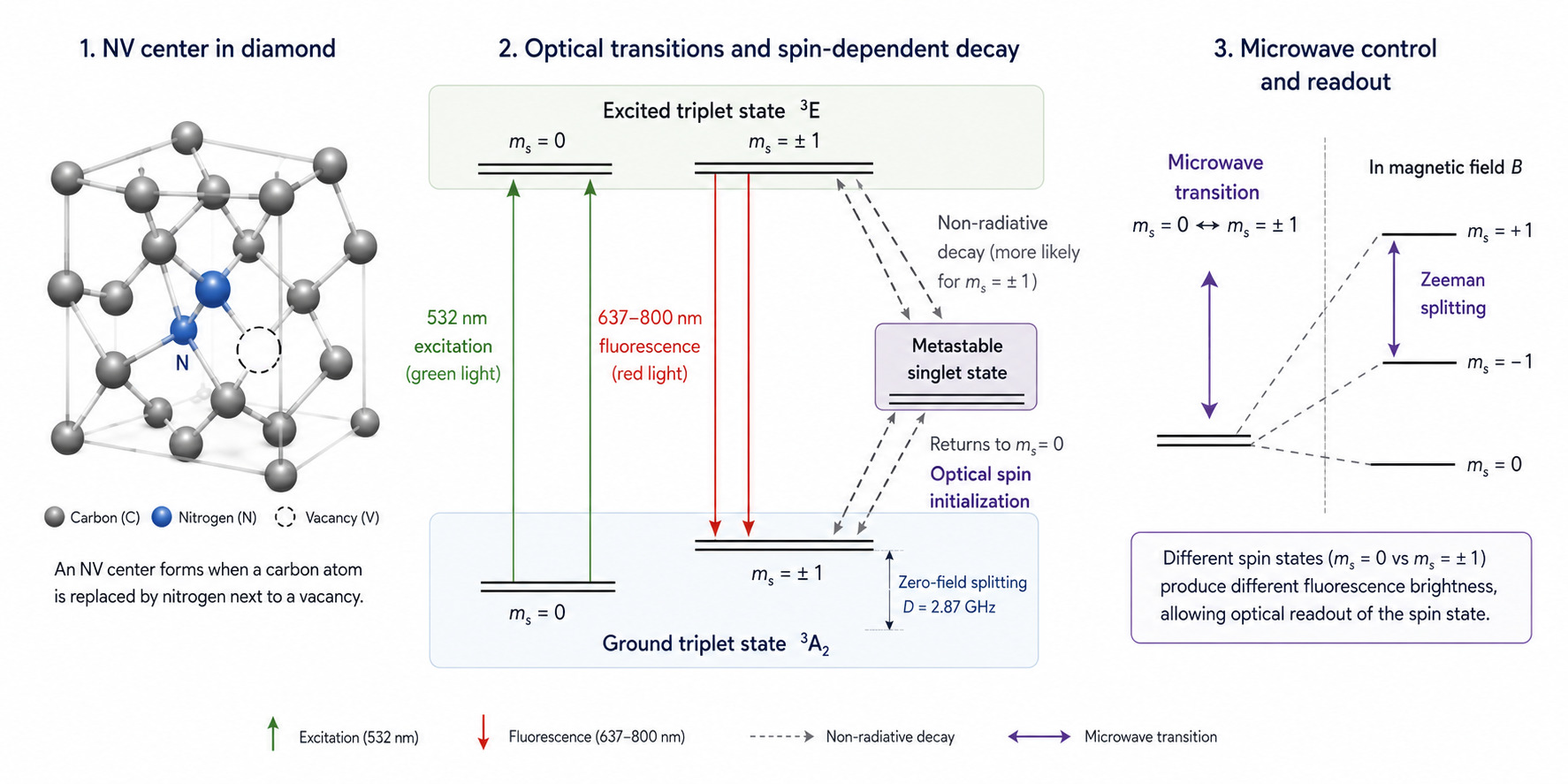
What exactly is an NV center? It is a point defect in the crystalline carbon lattice of a diamond. It forms when a single carbon atom is replaced by a nitrogen atom, and an immediately adjacent site in the lattice is left empty—creating a vacancy [5]. This system behaves like an isolated atom locked within a solid-state diamond matrix. It possesses its own distinct energy levels and its own atomic magnetic moment, known as spin, which can be easily initialized and read using ordinary light [3, 5] (Fig. 3).
Communicating with the Quantum World: The Principle of ODMR
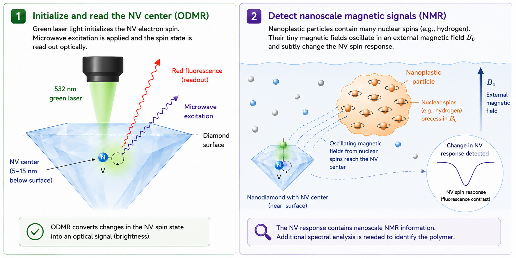
The remarkable feature of NV centers is that their quantum spin state can be manipulated and read using ordinary light at room temperature. When the diamond is illuminated with a green laser, the electrons within the defect are excited to a higher energy state. As they relax back to the ground state, they emit red photoluminescence, which can be easily measured.
The core principle relies on the NV center's atomic spin. To interact with this spin, the diamond must be prompted not only by the laser but also by invisible microwave radiation (similar to the frequencies used by mobile networks, but at a fraction of the power) [3, 5]. This microwave excitation acts like a fine radio tuner: it sweeps across frequencies to interact with the spins and prepare them for the measurement.
When an external magnetic field is applied, it shifts the spin energy levels of the NV centers within the diamond sensor. When the system is scanned with microwaves at this shifted resonance frequency, the electron spins are flipped, forcing them to relax through an alternative energy route that bypasses the direct path, causing the red light to dim slightly [2, 3, 5] (Fig. 4).
The magnitude of this fluorescence decrease depends strongly on the experimental setup. Early measurements on a small ensemble of NV centers showed a decrease of about 10% [2], whereas the optical contrast between the spin states of a single NV center can approach 30% [3]. This method of reading the magnetic state of the environment through changes in fluorescence intensity and tuned microwaves is called optically detected magnetic resonance (ODMR) [3, 5]. By precisely measuring how the microwave resonance frequencies shift in response to an external magnetic field, scientists can determine the field’s strength with high sensitivity.
Nanoscale NMR: A Chemical Fingerprint
This principle of brightness dips also extends to the detection of specific polymers. Common plastics, such as polystyrene and polyethylene, contain large numbers of hydrogen atoms. The nuclei of these atoms possess their own tiny nuclear spin and precess like microscopic spinning tops at a characteristic frequency (known as the Larmor frequency) when exposed to a magnetic field.
When a nanoplastic particle approaches the diamond surface within a distance of less than 20 nanometers, the rotating magnetic field of the plastic's hydrogen nuclei gently perturbs the spin of the nearby NV center [3, 6]. By applying rapid sequences of microwave pulses, scientists can filter out the chaotic environmental noise and isolate only the spin changes that match the specific precession frequency of hydrogen. By monitoring the resulting light dips of the diamond, researchers can perform nuclear magnetic resonance (nano-NMR) on a volume of just a few cubic nanometers. However, because hydrogen is found in almost all organic matter—including living cells—a simple hydrogen signal is not enough. Distinguishing a plastic particle from the surrounding tissue requires analyzing additional atomic details, meaning that a direct, live-ready nanoplastic detector is still a future research goal [6, 7] (Fig. 4).
Growing Quantum-Grade Diamonds
To ensure predictable results, researchers avoid unpredictable natural diamonds. Instead, high-purity synthetic crystals are grown in laboratories using Chemical Vapor Deposition (CVD) [5]. A vacuum chamber is filled with a mixture of hydrogen and methane gases, and microwave energy ignites a brilliant, high-temperature plasma. Carbon atoms precipitate from this plasma and deposit onto a substrate layer by layer. To eliminate background magnetic noise from the lattice itself, isotopically pure methane containing exclusively carbon-12 is used, as carbon-12 possesses no nuclear spin [3]. In the final stage of growth, trace amounts of nitrogen are introduced, and the crystal is subsequently irradiated with electrons and annealed to create a controlled distribution of stable NV centers localized just a few nanometers beneath the surface [5].
Current Bottlenecks and Outstanding Questions
Despite its immense promise, the technology faces significant hurdles. For an NV center to capture the incredibly weak magnetic fields of a nanoplastic particle, it often needs to lie only about 5 to 15 nanometers below the surface. However, the diamond surface is inherently unstable, plagued by dangling chemical bonds and surface contaminants that generate severe electrical and magnetic noise. This noise severely shortens the coherence time—the duration during which the NV center can maintain its quantum information—and limits the sensor's overall sensitivity [8].
Furthermore, the scientific community is currently grappling with conflicting experimental data.
Different surface treatments—including oxygen-, fluorine-, hydrogen-, and nitrogen-based terminations—can affect the charge stability and coherence of shallow NV centers in different ways. Their performance depends on the crystal surface, preparation procedure, and sensing conditions, so no single treatment is universally optimal [7, 8].
Liquid samples introduce additional complications at the complex diamond-water interface, where localized electrostatic fluctuations and ion adsorption can unexpectedly shift the resonance frequencies of shallow NV centers. Untangling these environmental effects from the actual plastic signal requires rigorous calibration and blank measurements, and remains a critical area for further investigation [6, 7, 8, 9].
From Living Cells to Field Microchips: Biological Challenges and Technological Visions
Detecting nanoplastics directly within the human body remains an urgent frontier for modern science. Due to their unique physicochemical properties and surface charges, experimental studies suggest that these minute particles can interact with biological barriers and, under certain conditions, may cross them—including the intestinal, placental, and blood-brain barriers, though the exact extent of this in humans is still being mapped [10, 11].
To study how nanoplastic particles influence cell processes, researchers synthesize model polymer particles with fluorescent nanodiamonds (FNDs) embedded directly inside them [10]. While the plastic matrix mimics the pollutant, the embedded diamonds serve as a tracer, showing so far low cytotoxicity, allowing scientists to reliably measure the cell's genuine response to the plastic itself without signal degradation [10].
Furthermore, the internalization of plastics often triggers a cellular immune response accompanied by a massive surge of free radicals, a state known as oxidative stress [12]. Because these radicals possess unpaired electrons, they behave like highly reactive miniature magnets, creating a localized magnetic unrest that nearby NV centers can detect. Since magnetic fields decay rapidly with distance, this sensing relies on nanodiamonds located at the particle’s outermost boundary or those gradually exposed as the polymer matrix undergoes cellular weathering. In a healthy, undisturbed cell, the NV center retains its quantum state for a relatively long period. While NV centers have been used to measure free radicals in human cells, and plastics are known to cause oxidative stress, combining these two steps into a single experiment has not yet been demonstrated. If successful, this method would provide an indirect but highly valuable indicator of cellular distress, helping toxicologists observe how cells react to plastic pollution [11, 12].
Moving this technology out of the controlled environment of a laboratory microscope and into natural water bodies or wastewater facilities introduces severe engineering complications. Natural water is highly heterogeneous, filled with background noise from dissolved iron or manganese ions and organic detritus that would completely blind a basic quantum sensor. To bypass this issue, the diamond sensor must be paired with dynamic decoupling protocols. By applying microwave pulses in a meticulously timed rhythm, the sensor acts as a temporal sieve. The slow, chaotic magnetic fluctuations of the environmental noise are actively canceled out, while the rapid, coherent precession of the hydrogen nuclei within the nanoplastics is amplified and isolated [6, 7]. Even with this quantum filtering, a sensor placed directly into a river or wastewater plant cannot do the job alone. Real-world analysis will still require traditional sample preparation, such as pre-filtering and calibration, to protect the diamond from fouling and ensure reliable measurements [3, 9].
Real environmental samples rarely contain a single pure polymer. They include mixtures of polyethylene, polystyrene, nylon, additives, natural organic matter, and weathered particles whose spectra may overlap. Vibrational-spectroscopy studies have therefore begun combining spectral measurements with machine-learning methods, including convolutional neural networks, to classify polymers and estimate weathering [1]. Similar computational tools might eventually assist in interpreting quantum-sensing data, but this remains a forward-looking proposal rather than a demonstrated nanoplastic application.
A further challenge is miniaturizing the optical, microwave, fluidic, and detection components. Current research has demonstrated microfluidic quantum-sensing platforms and NV measurements in flowing microdroplets [9]. Integrated light sources, microwave structures, and photodetectors are also being developed for diamond quantum technologies, but a portable, nanoplastic-specific quantum system-on-a-chip has not yet been demonstrated. A realistic intermediate step may be a stationary, automated instrument operating in a controlled laboratory or water-treatment workflow. Large-scale environmental mapping with such systems remains a long-term technological vision [4] (Fig. 5).
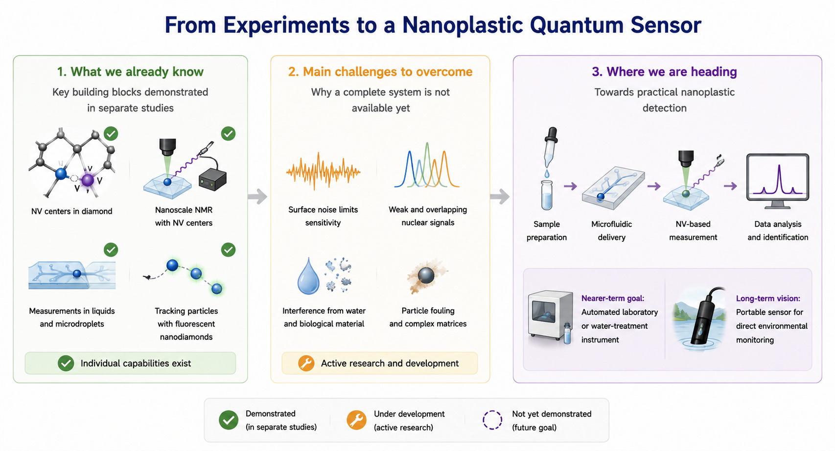
Quantum metrology leveraging nitrogen-vacancy centers in diamond offers a compelling technological perspective on how science might eventually probe the otherwise invisible domain of nanoplastics. While still largely in the developmental and laboratory stages, this approach highlights a potential pathway to bypass the diffraction limits of conventional optics and to gather indirect data from within complex environments such as living cells or fluid dynamics. Individual parts of this puzzle—like nano-NMR or cellular radical sensing—have already been proven to work separately. What hasn't been done yet is combining them into a single, direct, and ready-to-use plastic detector. If current engineering and surface noise challenges are successfully overcome, this fusion of quantum tech and computer science will eventually complement—not replace—conventional methods, giving us a powerful tool to study these elusive pollutants in targeted environmental and medical research.
About the authors

References
- Ivleva N. P. “Chemical Analysis of Microplastics and Nanoplastics: Challenges, Advanced Methods, and Perspectives.” Chemical Reviews 121 (2021): 11886–11936. https://doi.org/10.1021/acs.chemrev.1c00178
- Gruber A., Dräbenstedt A., Tietz C., et al. “Scanning Confocal Optical Microscopy and Magnetic Resonance on Single Defect Centers.” Science 276 (1997): 2012–2014. https://doi.org/10.1126/science.276.5321.2012
- Schirhagl R., Chang K., Loretz M., et al. “Nitrogen-Vacancy Centers in Diamond: Nanoscale Sensors for Physics and Biology.” Annual Review of Physical Chemistry 65 (2014): 83–105. https://doi.org/10.1146/annurev-physchem-040513-103659
- Centre for Quantum and Society. “Quantum for Good Challenge Launches Ahead of the UN International Year of Quantum: Harnessing Quantum Technologies to Tackle Global Challenges, with a Focus on Microplastics.” Quantum for Good, November 27, 2024. https://quantumforgood.eu/projects/challenge/
- Doherty M. W., Manson N. B., Delaney P., et al. “The Nitrogen-Vacancy Colour Centre in Diamond.” Physics Reports 528 (2013): 1–45. https://doi.org/10.1016/j.physrep.2013.02.001
- Allert R. D., Briegel K. D., Bucher D. B. “Advances in Nano- and Microscale NMR Spectroscopy Using Diamond Quantum Sensors.” Chemical Communications 58 (2022): 8165–8181. https://doi.org/10.1039/D2CC01546C
- Janitz E., Herb K., Völker L. A., et al. “Diamond Surface Engineering for Molecular Sensing with Nitrogen-Vacancy Centers.” Journal of Materials Chemistry C 10 (2022): 13533–13569. https://doi.org/10.1039/D2TC01258H
- Sangtawesin S., Dwyer B. L., Srinivasan S., et al. “Origins of Diamond Surface Noise Probed by Correlating Single-Spin Measurements with Surface Spectroscopy.” Physical Review X 9 (2019): 031052. https://doi.org/10.1103/PhysRevX.9.031052
- Sarkar A., Jones Z. R., Parashar M., et al. “High-Precision Chemical Quantum Sensing in Flowing Monodisperse Microdroplets.” Science Advances 10 (2024): eadp4033. https://doi.org/10.1126/sciadv.adp4033
- Li R., Vedelaar T. A., Sigaeva A., et al. “Fluorescent Nanodiamonds for Tracking Single Polymer Particles in Cells and Tissues.” Analytical Chemistry 95 (2023): 13046–13054. https://doi.org/10.1021/acs.analchem.3c01452
- Ahn J., Hronová K., Pashigreva A., et al. Nanoplastics: A Systematic Risk Analysis for Human Health, Ecosystems, and the Environment. ALLATRA Global Research Center, 2026. https://doi.org/10.65849/agrc.report.mnp.2026.04001
- Nie L., Nusantara A. C., Damle V. G., et al. “Quantum Sensing of Free Radicals in Primary Human Dendritic Cells.” Nano Letters 22 (2022): 1818–1825. https://doi.org/10.1021/acs.nanolett.1c03021
© 2026 Tatiana Kárová. This article is published under a Creative Commons Attribution 4.0 International licence (CC BY 4.0) unless a different licence is stated in the credit line of an individual item. The authors retain copyright; ALLATRA Natural Science Review holds the right of first publication. The Review charges no submission, publication, or article processing fees. © 2026 ALLATRA Inc.